What is Pineal Gland Tumor?
Pineal Gland Tumor is a neoplasm that arises from the cells in pineal gland. These tumors are extremely rare. They make up 0.4-1% of all intra-cranial tumors in adults, and 3-8% of brain tumors in children. There are some tumors that can arise in the area of pineal gland, but these tumors require a different approach [2].
For information on other intracranial tumors read: Rhabdoid tumor, Diffuse intrinsic pontine glioma, craniopharyngioma.
Pineal gland
Pineal gland is a small endocrine organ, which has a shape that represents a pine cone. It is about 0.8 cm long and located in the rear end of third ventricle- a fluid filled cavity in the brain. It is composed of pineal cells. With age calcium, fluoride and phosphorus build up in the gland, making it look calcified in X-rays. Pineal gland produces melatonin- hormone which plays the most important role in regulating human circadian cycle (biological clock). Melatonin also helps to regulate some of the reproductive hormones. Still, the exact mechanisms of how melatonin works in the human body are not understood.
- Circadian rhythm- melatonin secretion is stopped by the daylight, but during night it is high
- Reproduction- pineal hormone blocks secretion of luteinizing and follicle stimulating hormone [1].
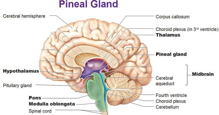
Anatomical location of pineal gland.
Image source: upliftconnect.com
Causes and types
The exact cause of pineal tumor is not known. Tumors in pineal gland can arise from different types of cells:
- Germ cell tumors account for more than 50% of pineal tumor cases. These tumors usually have distinct boundaries, but it is characteristic for these tumors to seed in the ventricles that contain cerebrospinal fluid. There are different types of germ cell tumors:
- Embryonal cell carcinoma-highly malignant
- Endodermal tumor -very invasive
- Teratomas– well formed, round, multicystic and lobuated. These tumors usually compress surrounding structures.
- Parenchymal tumors- tumors formed from functional tissue. Account for 20-30% of all pineal gland tumors.
- Pineoblastoma
- Pineocytoma
- Other types of pineal tumors:
- Astrocytoma
- Meningioma
- Metastatic tumor
- Lymphoma
- Pineal cyst[4]
Symptoms
Due to location of the tumor, they often present with hydrocephalus- fluid pressure buildup in the brain. The fluid buildup causes ventricles to expand and put pressure on other brain structures. This causes symptoms like:
- Nausea and vomiting
- Headaches
- Seizures
- Memory disturbance
- Changes in vision
- Life threatening brain herniation
If the nearby tectal region is involved, various eye problems can arise. This region is responsible for visual reflexes. Symptoms include:
- Inability to focus on an object
- Double vision
- Eye movement impairment- inability to look upward
- Nystagmus
Other symptoms:
- If the tumor invades thalamus, it is possible to lose sensation and motor function in one half of the body.
- Involvement of hypothalamus can cause impairment of thermal and fluid regulation, and cause sleepiness and weight gain. Also read: pituitary gland tumor.
- Tremors
- Lack of coordination
- Hearing impairment- buzzing, ringing sound
- Drowsiness
- Changes in speech
- Behavior changes[2,3]
Diagnosis
Physical examination
A lot of initial symptoms are unspecific and can be explained with other conditions. A thorough patient history is necessary. To diagnose pineal tumor, doctor will need to do a thorough physical examination, where neurological examination plays the most important role. It is important to check:
- Vision
- Hearing
- Balance and coordination
- Reflexes
After physical examination, if brain tumor is suspected, doctor can order further investigations [2,3].
Imaging studies
- X-Ray imaging– can detect highly calcified pineal gland, but is not enough to make a diagnosis
- CT scanning– can be helpful in detecting level of hydrocephalus and calcification of the gland. Also used for guiding during biopsy.
- MR imaging– method of choice for diagnosing pineal gland tumor. Usually high resolution MRI with gadolinium is used. Tumor size, blood supply, homogeneity and involvement of surrounding structures can be assessed [2,3].
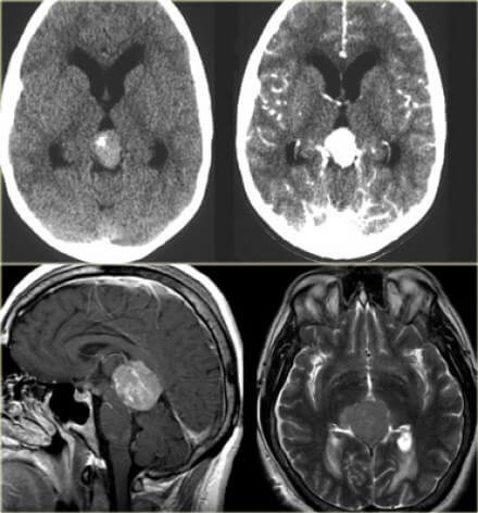
Pineal gland tumor in CT and MRI scans.
Image source: radiologyasisstant.nl
Laboratory studies
Cerebrospinal fluid and blood serum marker detection can be helpful in diagnosing pineal gland tumors. Tumors contain some molecular characteristics from their initial cell lineage. Detecting the embryonic proteins can be helpful in diagnosing the type of tumor. CSF and serum testing is used for diagnosis and also for monitoring the response to treatment [2,3].
Biopsy
Biopsy is necessary to determine the type of tumor, which allows deciding on the treatment options[2,3].
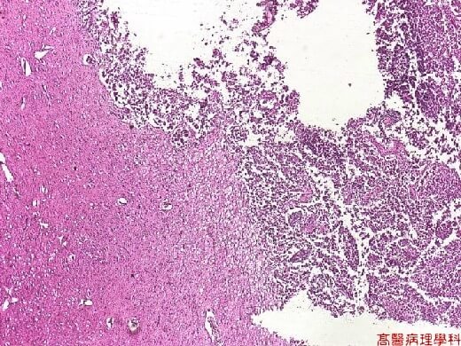
Histology image of pineal gland tumor which extends into third ventricle.
Image source: http://pathology.class.kmu.edu.tw/
Treatment
Surgical removal
Surgical removal of pineal tumor is one of the most difficult neurological surgeries, due to the location of the pineal gland. In some cases surgery can be curative, but in other cases it can only reduce the size of the tumor and provide a definitive diagnosis. Usually minimally invasive surgery is used.
Surgery is also used to reduce the CSF buildup in the ventricles. Usually an endoscopic third ventriculostomy is used- creating a hole for drainage. In other cases a shunt is used [4,5].
Radiation therapy
In most cases, pineal gland tumors are sensitive to radiation therapy. Nowadays targeted radiation therapy is often used. In children, radiation therapy has significant effect on cognitive development. In both children and adults radiation therapy is associated with high mortality rates [4,5].
Chemotherapy
Chemotherapy is often used together with radiation therapy. The choice depends on the type of tumor and general condition of the patient. Chemotherapy can actually minimize the need for radiation [4,5].
Prognosis
The prognosis highly depends on the type of tumor. Usually germ cell tumors have excellent prognosis, because they are responsive to radiation and chemotherapy. Non germ cell tumors have a worse prognosis. Surgical removal of the tumor also has its risks, because the tumor is very hard to access.
Radiation therapy can cause complications, such as:
- Cerebral necrosis
- Secondary tumors (like meningioma)
- Endocrine system dysfunction[6]
If you found this article helpful, share it on social media. For your comments and personal experience use the comments section below.
References
- Pineal gland: https://www.endocrineweb.com/endocrinology/overview-pineal-gland
- General information for patients: http://www.emedicinehealth.com/pineal_tumors/article_em.htm
- Detailed information for health professionals: http://emedicine.medscape.com/article/249945-overview#a7
- Types of tumor, treatment: http://neurosurgery.ucla.edu/pineal-tumor
- Treatment: http://www.cancerresearchuk.org/about-cancer/type/brain-tumour/treatment/types/treatment-for-pineal-region-tumours
- Prognosis: https://www.ncbi.nlm.nih.gov/pubmed/9548342
Similar Posts:
- Pituitary Gland Tumor
- Pineoblastoma
- Inoperable Brain Tumor
- Brenner Tumor
- PNET Tumor
- Retinoblastoma
- Ovarian Teratoma


Leave a Reply