What is Brenner tumor?
Brenner tumor is a rare form of ovary tumors. These tumors belong to a group called epithelial tumors (also see Nasopharyngeal carcinoma). Only approximately 2% of all ovarian tumors are Brenner tumors. There are three types of Brenner tumor:
- Benign
- Borderline
- Malignant[1]
Types of Brenner tumor
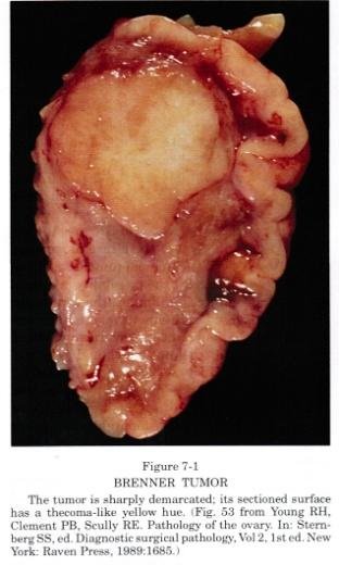
Benign Brenner tumor
Benign Brenner tumor is derived from normal surface epithelium of the ovary. This tumor is usually found in one ovary, but in 6% of cases it can be in both ovaries. Benign tumor has a firm consistency and yellow coloring. Microscopically benign Brenner tumor is characterized by: 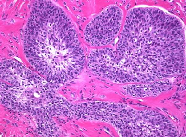
- Solid, cystic nests of mature cells that resemble transitional epithelium (like the one found in bladder)
- Cell surrounding stroma is dense and fibrous
- Clear outline of the cells

- Cytoplasm is pale, and cells have small nucleoli
- In epithelial nests microscopic cysts can be seen[2]
Borderline Brenner tumor 
Borderline Brenner tumors are also called atypical proliferative tumors because they are mostly benign by nature, but the cells in the tumor have a higher proliferation rate than benign tumor. These tumors are more seen in women around 60 years of age. Tumor size is usually larger than benign, and some symptoms can be present due to its size. These tumors are associated with urothelial carcinoma of the bladder. 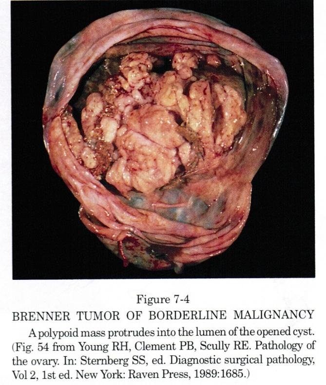
These tumors are usually present on one side, and contain cysts in various locations of the ovary. The mass protrudes in the lumen. Microscopically the tumor is characterized by:
![]()

- Solid and papillary proliferative epithelium
- Nuclei can be moderately atypical
- Papillary areas that protrude in the lumen can have multilayer epithelium with prominent nucleoli
- No stromal invasion [3]

Malignant Brenner tumor
Malignant Brenner tumor is very rare. These tumors resemble trasitional cell carcinoma of the ovary and it is very important to differentiate these two types of tumor, because the latter has a poor prognosis. Malignant Brenner tumor is usually unilateral, but they can affect both ovaries. They can have metastasis in the pelvic area, but metastases can also be found in distant lymph nodes, lungs, liver and bones. Microscopically this tumor is characterized by:
- Atypical cells
- Stromal invasion
- Consists of transitional, squamous or undifferentiated cells or mixture of them
- High recurrence rate [4]


![]() Brenner tumor clinical presentation
Brenner tumor clinical presentation
Symptoms
Brenner tumor is usually asymptomatic. Benign tumors rarely cause any symptoms. It can sometimes cause discomfort or pain in lower abdomen. Mostly Brenner tumors are an incidental finding during ultrasound examination or pelvic surgery.
Associated diseases
- Endometrial adenocarcinoma (also see Vaginal adenocarcinoma)
- Postmenstrual bleeding
- Endometrial hyperplasia
- Transitional tumor of the bladder[1]
Brenner tumor diagnosis
Laboratory findings
There are usually no specific findings in blood analysis for Brenner tumor. In some cases tumor marker CA-125 can be elevated, but this finding is very unspecific. It can be positive in other types of cancer, for example Fallopian tube cancer.
The diagnosis is usually made after getting a sample of the tumor via laparoscopy or biopsy. Detailed histological description- see section “Types of Brenner tumor”.
There are criteria for diagnosing malignant Brenner tumor, and these are:
- Malignant histological features
- Stromal invasion by elements of epithelium
- Malignant tumors usually do not arise from benign, but there should be an association between malignant element and benign Brenner tumor
- Mucinous cystadenoma should be absent or well separated from benign and malignant Brenner tumor[5]
Immunohistochemistry
This method uses antibodies to find specific proteins associated with certain diseases. In case of Brenner tumor some specific proteins can be found. Since Brenner tumors have a lot of similarities with transitional cell tumors, immunohistochemistry can be helpful in makng the correct diagnosis.
Brenner tumors show lower immunoexpression for all markers. Borderline Brenner tumors strongly express marker EGRF, while it is usually negative in transitional cell tumor. Malignant Brenner tumors are positive for Cyclin D1, Ras and EGFR proteins, but Transitional cell tumors are negative immunoexpression for these markers [6].
Brenner tumor radiology
Ultrasound imaging is the method of choice for imaging of Brenner tumor. This investigation is painless and easy to perform. Usually Brenner tumors are incidental finding during gynecological investigation. Ultrasound findings can vary greatly depending on the type of tumor:
- Benign tumor: solid structure, in 50% of the cases there can be calcifications. The presentation is very unspecific therefore Brenner tumors can be confused with other ovarian neoplasm (see Germ cell cancer). In size they are usually 2- 6 cm, although there has been a case report on tumor reaching 39 cm in diameter. Benign tumors can be multilocular.
- Borderline tumor: usually larger than benign tumor, up to 9 cm in diameter. Mass is usually solid, located in one place. These tumors usually are not associated with fluid in Douglas cavity.
- Malignant tumor: these tumors can vary a lot in size and they have irregular walls. They are solid and often multilocular. In Dopler imaging moderate to high blood flow can be observed. Calcifications can be present. Fluid in Douglas cavity can be found [7].
MRI
MR imaging can also show Brenner tumors. Usually it is an incidental finding. On MRI Brenner tumors have a solid, hypointense appearance and they can be confused with fibroma. Some Brenner tumors have a cystic component but they usually do not contain fluid. Large Brenner tumors (>10 cm) have no signs of ascitis, local invasion, lymph node invasion or metastases in other organs [8].
Brenner tumor treatment
The method of choice for treating Brenner tumors is surgery. Benign and borderline tumors have a low rate of recurrence after surgery. Malignant tumors have a high rate of recurrence. For patients with malignant Brenner tumor, chemotherapy can be used after surgery [1].
If you find this article helpful, share it on social media.
Use the comments box below for your personal experience and other inquiries.
References
- Patient information about Brenner tumor: https://rarediseases.info.nih.gov/diseases/9397/brenner-tumor-of-ovary
- Benign Brenner tumor: http://www.pathologyoutlines.com/topic/ovarytumorb9brenner.html
- Borderline Brenner tumor: http://www.pathologyoutlines.com/topic/ovarytumorborderlinebrenner.html
- Malignant Brenner tumor: http://www.pathologyoutlines.com/topic/ovarytumormalignantbrenner.html
- Laboratory tests for Brenner tumor: http://www.ccij-online.org/article.asp?issn=2278-0513;year=2015;volume=4;issue=4;spage=584;epage=586;aulast=Sangwaiya
- Diagnostics of Brenner tumor: https://www.ncbi.nlm.nih.gov/pubmed/19033864
- Imaging of Brenner tumor: http://onlinelibrary.wiley.com/doi/10.1002/uog.11149/full
- Imaging of Brenner tumor: http://www.jultrasoundmed.org/content/25/10/1245.full
Similar Posts:
- Serous Cystadenoma
- Ovarian Teratoma
- Krukenberg Tumor
- HER2 Positive Breast Cancer
- Urethral Cancer
- Granular Cell Tumor
- DCIS Breast Cancer


Leave a Reply