What is Dermatofibroma?
Dermatofibroma, otherwise known as benign fibrous histiocytomas, dermal dendrocytoma, Fibrous dermatofibroma, Fibrous histiocytoma, Nodular subepidermal fibrosis, and Sclerosing hemangioma, is a skin growth, considered benign. It is found mainly on the legs and can grow up to 1cm in diameter. The condition primarily consists of fibrous tissue or scar like tissue under the skin.
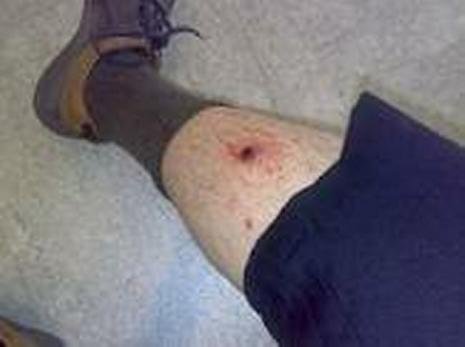
They occur as single lumps or as multiple lumps. The skin growth is usually round and brownish to purple in color and feels like a hard lump in the skin. Dermatofibroma is also called fibrous histiocytoma because there is a growth of dendritic histiocyte cells that are normally non-cancerous. However, some cases can develop malignant carcinomas.
Researchers believe that these skin growths are due to a reaction to previous injuries. Dermatofibroma has a persistent nature which makes other researchers believe that it is a neoplastic process rather than a reaction.
Women have a higher risk of developing dermatofibroma. The condition is usually asymptomatic, but others may experience pruritus (severe itching) and tenderness.
Dermatofibroma Causes
The exact cause and mechanism of dermatofibroma is unknown although some risk factors have been identified such as:
- Injury – Previous skin injuries such as thorn pricks or insect bites may lead to dermatofibroma. Injury to the skin stimulates the cells to regenerate.
- Gender – Women are more prone to develop the skin condition than men. Female to male ratio is 4:1.
- Heredity – Family history of dermatofibroma contributes to its development
- Age – Dermatofibroma usually develops in young and middle aged adults.
Dermatofibroma Symptoms
Symptoms of dermatofibroma include:
- Small, round skin growth on the skin, about 1cm in size
- Red, brownish to tan or purple in color, may change in color over time
- May have a scaly and shiny surface
- Usually appears on the legs, but it can also develop on the arms, face and trunk
- Dimples inward when pinched or squeezed together
- May bleed if irritated or damaged
- Painless, but may be tender upon touching
- Sometimes itchy
- May appear as a single bump or multiple bumps
- Develops gradually over several months
Dermatofibroma Diagnosis
Appearance of any skin growths requires early diagnosis to rule out the presence of malignancies. A change in color of the skin growth usually entails skin cancer formation. However, ruling out malignancies is only done because some symptoms of dermatofibromas are similar to that of other skin malignancies. Tests for dermatofibroma usually reveal a benign condition. Diagnostic tests and exams include:
Physical examination -Dermatofibromas are initially diagnosed by physical examination of the skin and the growth itself. The physician usually squeezes the lump and dimpling occurs. This indicates dermatofibroma.
Histological Examinations – When other symptoms occur such as, bleeding or change in color, a sample of the tissue is excised for biopsy purposes. The lump is examined using a microscope to determine the characteristics of the cells and if they are benign or malignant.
Histological examinations may reveal a pseudoepethiliomatous hyperplasia which means there is a false epithelial growth on the skin. This may result from fibroblast actions over the keratinocytes of the injured skin.
Basal cell carcinomas (skin cancer) have been reported in dermatofibromas. Due to this, it is important to seek medical advice whenever an abnormal skin growth appears. Although dermatofibromas are benign, a potential for malignancy can still arise.
Dermoscopy – Dermoscopy is an adjunctive diagnostic procedure for dermatofibroma. The findings usually present a peripheral pigment network with a whitish central area.
Dermatofibroma Treatment
Dermatofibromas do not necessarily require any treatment. Being normally benign, it can be left in place and ignored, but reassurance is important for patients. However, for aesthetic purposes, particularly for female patients, there are surgical or medical treatments to remove the lesion.
- Medical Care – Medical treatment only includes steroid injections to relieve any symptoms such as pain and itchiness. It also decreases the tendency for the lump to increase in size.
- Surgical Care – For disturbing dermatofibromas, such as in cases of pain and pruritus, the skin growth may be removed surgically. Presence of indefinite histologic examinations may also require this in order to remove any cells that may be malignant. Surgical treatment involves various techniques such as:
- Excision Biopsy – Surgical removal involves excision of the entire skin growth including the subcutaneous layer underlying it. Inverted pyramidal biopsy is usually done to reduce scar formation.
- Shaving – Shaving the skin may be done for cosmetic purposes. This is done by superficially removing the lump with the use of a surgical knife. However, this procedure increases recurrences because the lesion is not removed from within.
- Cryosurgery – This involves the exposure of the skin growth to very cold temperature such as liquid nitrogen to freeze and remove the lump. Just like superficial shaving, the lump may also reoccur and may require another treatment for removal.
- Laser Treatment – Carbon dioxide laser treatment can be done and has been reported to effectively remove the skin growth.
Dermatofibroma Home Remedies
Aside from medical and surgical treatment, dermatofibromas can be treated by home remedies such as:
- Apply spirit of camphor over the area. Apply continuously until the skin dries up. This procedure may cause stinging of the skin.
- Use milk of magnesia. Apply the solution for 10 minutes and rinse off.
- Increase intake of low-fat, high fiber diet. Fruits and vegetables are essential in improving the skin texture.
- Apply cellulite cream. Cellulite creams also improve the appearance of the lump.
- Wash face with Benzotn solution. Mix two to three drops of Benzotn solution in a glass of cold water. Use this for washing the face. It helps prevent infection of the lesion because it contains antibacterial properties.
- Avoid too much exposure to the sun during periods from 10am to 3pm because exposure to ultraviolet rays may worsen the condition and trigger a malignancy.
Any change in the features of a dermatofibroma should be reported to health practitioners. Dermatofibromas mimic skin malignancies, so early treatment and management is essential.
Pictures
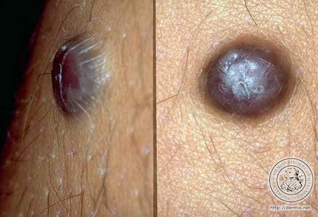
Source – dermis.net
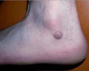
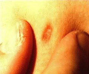
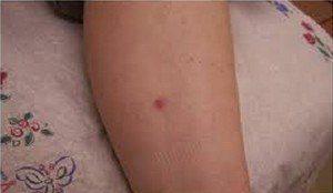
Updated by Dr. Kristine on 26/8/2012
Similar Posts:
- Solitary Fibrous Tumor
- Duodenal Cancer – Symptoms, Prognosis, Survival Rate and Treatment
- Syringoma – Treatment, Removal, Pictures, Causes, Surgery, Prevention
- Mast Cell Tumor
- Amelanotic Melanoma – Pictures, Symptoms, Prognosis
- Epithelioid Sarcoma
- Squamous Cell Carcinoma in Situ – Pictures, Treatment, Symptoms



Leave a Reply