What is Myxoma?
Myxoma is a benign tumour of mucous tissue, a type of primitive connective tissue. Mucous tumours have a specific consistency that resembles an umbilical cord and pathological appearance: they are globule-like, sometimes on a peduncle or sessile with wide attachment on the base.
The tumour is gelatinous, white or yellowish, and may grow up to 15 cm. Histologically, they are composed of fibroblast cells of the connective tissue that are submerged in the gelatinous, mucous tissue.
Myxomas appear more frequently in women, for unknown reason. These tumours may appear sporadically, or appear in form of inherited conditions. Only 10% of myxomas are genetically influenced.
One of the inherited syndromes that include myxomatous tumours is Carney complex, where there are multiple myxomatous tumours, skin hyperpigmentation due to adrenal hyperfunction, schwannomas, testicular tumours and pituitary adenoma with production of growth hormones.
This syndrome is inherited autosomal dominantly. Another syndrome is referred to as Mazabraud syndrome. It is a condition where myxomatous tumours appeared associated with McCune-Albright syndrome. The myxomas appear on skin, heart and in bones. (1)
Types
There are several types of myxoma, depending on the organ where they develop, and their aggressiveness. Types are:
- Cutaneous myxoma (on skin or subcutaneous tissue, usually in Carney complex syndrome),
- Atrial heart myxoma (the most common primary heart tumour, usually located in the left atrium),
- Intramuscular myxoma (in sceletal muscles; it is difficult to diagnose, because it is very rare),
- Juxta-articular myxoma (located in the joints, shoulder, ankle or hip),
- Digital myxoma (located at the tip of the fingers),
- Aggressive angiomyxoma (forms in the body cavities, vulvovaginally, or in the peritoneum),
- Angiomyofibroblastoma (well marginated and may appear on skin or vulva),
- Nerve sheath myxoma (develops at the layers of the nerve and most commonly, they appear on the legs, knees),
- Pseudomyxoma peritonei (this tumour may be aggressive, and spreads quickly over the peritoneum to different organs). (2)
Symptoms
Signs and symptoms are different depending on the location of the tumour. They may be unnoticed if they grow in the inner organs or muscles. If the myxoma is in a skeletal muscle, the only symptoms are a presence of a growing mass without any pain and possible hormone problems. The possible locations of this tumour are upper leg and shoulder and upper part of the back.
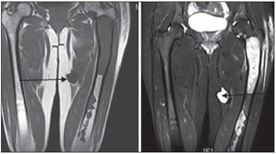
Figure 1 Intramuscular myxoma on left thigh (Mazabraus syndrome) (3)
If the myxoma is in the heart, it is usually with a peduncle, and located in either right or left (more often) atrium. These tumours if small, won’t develop any symptoms in a person, but as they grow, they may induce problems with blood flow, valvular problems (stenosis or regurgitation), arrhythmias and embolism in the heart or if they detach, the particles of tumour may travel to other organs and deprive their circulation.
That is how the kidneys, brain and other organs develop ischaemia and a person may suffer from stroke. If the presence of myxoma is long enough it causes structural and functional changes in the heart causing its failure. The symptoms of heart failure depend on the side where the myxoma is. If on right atrium the symptoms would include swelling in the body, peripheral oedema and abdominal distension. If the myxoma is in left atrium, the symptoms include dizziness, trouble with breathing, coughing and haemoptysis, coughing of blood, but also frequent respiratory infections, cyanosis, weight loss, difficulty sleeping on bed without pillow, syncope etc. (4)
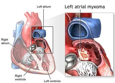
Figure 2 A large myxoma of the left atrium (5)
Myxoma on fingers doesn’t have much place to spread and grow which is why this myxoma induces severe pain. Skin myxoma may be a part of Carney complex r not. It may appear as a growing mass on face (there were cases of myxoma present even in the external ear canal), trunk or extremities.
Some of these myxomas appear to be reddish, which speaks of presence of the angiomatous vascular component of tumour (angiomyxoma). Some of these tumours may appear on genitals, which is when the differential diagnosis needs to be done to exclude more aggressive type of myxoma, the aggressive angiomyxoma. (6)
Juxta-articular myxoma is mostly developed on the knee (88% of all juxta-articular myxomas), shoulder, elbow, ankle and hip. The symptoms may include pain in the joint and tenderness, difficulties with movements and swelling or may be an accidental finding on the X-ray. People with osteoarthritic changes in the joints or have previously injured a joint, have a higher risk of developing juxta-articular myxoma. (8)
Diagnosis
There are differences in diagnostic procedures for each type of a myxoma. Skin myxomas need histological verification and also some histo-immunological markers must be analysed to prove whether or not it is associated with Carney syndrome.
Histological analysis is performed after excision of the tumour. Specific findings include mucous matrix with a small number of fibroblasts and some specific stains on the microscopic slide: eosin, hematoxylin, vimentin, desmin, alcian blue etc.
Atrial myxoma is diagnosed with ultrasound, and more precise information are gathered with transesophageal ultrasound. The myxoma is visible in the cavity of left or right atrium.
Clinical exam is the first step of the evaluation of the patient which may bring the diagnosis closer, providing the possible location of the tumour. Auscultation of heart may bring some diagnostic dilemmas, which is when some further diagnostic procedures are ordered: ECG, ultrasound, MRI, CT. On X-rays, CT and MRI the tumour is presented as a solid, soft tissue mass, on a peduncle or with a wide attachment on the base of the cavity.
Treatment
These tumours are very fragile, and prone to fragmentation. That is why the surgery is performed with a caution. Surgical procedure is the only way to extract the tumour completely. The procedures bring different levels of risk, depending on the location. Sometimes if the extraction wasn’t complete there is a chance for a relapse.
Sometimes there is some use from medications, to treat pain, arrhythmias, heart failure etc.
The prognosis is good, and in most of the types there is no chance for metastasing. Some of the myxomas are aggressive, but when they have been treated aggressively, there is a chance for a complete recovery. (1)
Works Cited
- Myxoma. tumour surgery.org. [Online] [Cited: 3 12, 2017.] http://www.tumorsurgery.org/tumor-education/soft-tissue-tumors/soft-tissue-tumor-types/myxoma.aspx.
- Myxoma. Primehealthchanel. [Online] 2012. [Cited: 3 12, 2017.] http://www.primehealthchannel.com/myxoma.html.
- Mazabraud syndrome. Mary John A, Behera KK, Mathai T,Parmar H, Paul TV. 2013, Indian Journal of Endocrinology and Metabolism 17(4), pp. 740-2.
- GK., Sharma. Atrial myxoma. emedicine Medscape. [Online] 2017. [Cited: 3 11, 2017.] http://emedicine.medscape.com/article/151362-overview.
- Atrial myxoma. Dmarkusalanis. [Online] [Cited: 3 12, 2017.] http://drmarkusalanis.tumblr.com/page/12.
- Cutaneous myxoma. Fibrohistocytic tumours. [Online] [Cited: 3 12, 2017.] http://freecontent.lww.com/wp-content/uploads/2014/09/Requena-Ch13-Cutaneous-Myxoma.pdf.
- olitary superficial acral angiomyxoma: An infrequently reported soft tissue tumor. Kura MM, Jidal SR. 2014, Indian journal of dermatology 59(5), p. 529.
- Juxtrarticular myxoma. Dovemed. [Online] 2016. [Cited: 3 12, 2017.] http://www.dovemed.com/diseases-conditions/juxta-articular-myxoma/.
Similar Posts:
- Neuroendocrine Cancer – Symptoms, Treatment, Prognosis, Survival Rate
- Leiomyosarcoma – Survival Rate, Symptoms, Prognosis, Treatment
- Keratoacanthoma – Pictures, Treatment, Symptoms, Causes
- Adrenal Adenoma
- Syringoma – Treatment, Removal, Pictures, Causes, Surgery, Prevention
- Mast Cell Tumor
- Dermatofibroma


Leave a Reply