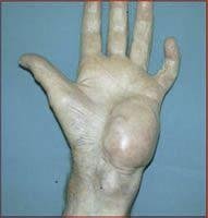
What Is Histiocytoma?
Histiocytoma is also known as fibrous histiocytoma. It is a type of tumor that occurs both in your bone and soft tissue. Histiocytoma can be cancerous or non-cancerous. Cancerous histiocytoma has high potential to cause cancer in soft tissues of your body such as limbs, legs and abdomen. A non-cancerous tumor is highly unlikely to cause cancer and occurs on your skin or bone. 1
Causes
Studies are yet to establish the exact causes of histiocytoma. However, cancerous histiocytoma is associated with cancer and various triggers of cancer are suspected to be its cause. They include:
Genetic factors
Defective genes are thought to cause cancerous histiocytoma. A person who comes from a family where cancerous tumors run in the family is more likely to develop cancerous histiocytoma.
Exposure to radiation
Radiation therapy, a method that uses high beam energy to destroy cancer cells can trigger malignant tumor in your body. Those people who undergo therapy that uses radiation are at a higher risk of developing cancerous histiocytoma.
Exposure to chemicals
Exposure to particular chemicals can elevate your risk of developing cancerous tumor. Doctors believe that chemicals such as arsenic and vinyl chloride can promote cancer, although studies are underway to confirm it
Symptoms
- Signs and symptoms of histiocytoma depend on whether the tumor is benign or malignant. For non-cancerous tumors, patients report of pain after an injury or fracture. The affected area can be tender without any swellings or other symptoms.
- Non-cancerous tumors can also affect your nervous system by compressing the spinal cord and the nerves. This can cause pain, memory loss and difficult in understanding speech as well as confusion.
- Malignant tumors in soft tissues can develop within three months without causing any pain or other symptoms. Once the tumor becomes big, it exerts pressure on your nerve causing pain. The following are symptoms of cancerous histiocytoma:
- Patients with cancerous histiocytoma feel pain when the tumor compresses the nerve or muscles. A patient also has limited motion in the leg or arm which can cause the person to limp while walking.
Diagnosis
A number of tests can be used to diagnose histiocytoma. These tests include:
Physical test
Your doctor would want to know your medical history and symptoms you are experiencing. Make sure you inform your doctor the medicines you have been using or any past or present medical condition. Your doctor will evaluate your symptoms and medical history to help him understand your condition.
Imaging tests
Your doctor can also use imaging techniques to scan the affected area and determine the problem. Your doctor can use magnetic resonance imaging which uses powerful magnetic waves to produce detailed pictures of soft tissue in the body.
The MRI scan helps the doctor check for any growths and abnormalities in the soft tissues. A computed tomography scan is used to create images of different sections of the affected areas to assist in diagnosis of this condition.
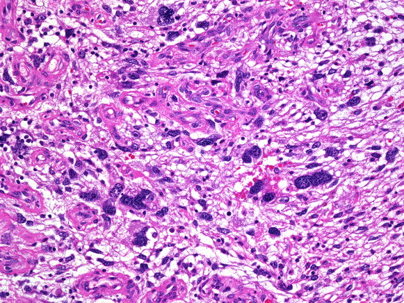
Biopsy
Your doctor can also conduct a biopsy to assist in diagnosing this condition. The doctor will remove a sample of your affected tissue and examine it under a microscope for any abnormalities. The sample is then taken for pathological test to find out if it is cancerous or not.
Additional tests are required to rule out other similar tumors such as synovial sarcoma, endometrial stromal sarcoma, myofibroma and myopericytoma etc.
Treatment
Histiocytoma can be managed depending on the type, size and location. Since these tumors cause pain, you will be given pain killers to relieve pain. Other treatment options for histiocytoma include:
Surgical excision
This procedure is necessary to remove skin tumor or lesion completely by using a cutting instrument such as scalpel. In this method, the doctor will inject anesthesia near the tumor to relieve pain during surgery and enlarge the tumor so that it is easily removed.4
Your doctor uses sharp tool to cut the tumor and remove it. When the tumor has been removed, the doctor can use dermal loop electrode to remove cells from the tumor that may have been left. This can also help reduce scarring by combing the margin of the wound with your nearby skin.
Bleeding is inevitable during surgery; your doctor will apply aluminum chloride hexahydrate and other chemicals on the affected area to prevent bleeding.
A relaxing antibiotic ointment is also applied at the place of surgery after cleaning to boost healing. The doctor covers the wound to prevent infections.
Radiation therapy
This procedure involves the use of powerful radiation to destroy cancer cells. It is also useful in reducing the risk of the cancerous tumor reappearing after surgery.
Your doctor can use any of the following direct radiation to the affected tissue:
- Your doctor can place a linear accelerator outside your body and direct strong beam to the tumor.
- The doctor can place a substance in your body that releases radiation to kill the tumor.
- Radiation therapy can be used together with hyperthermia. Hyperthermia therapy involves the use of microwave or ultrasound to heat cancerous tumors at high temperatures. Hyperthermia therapy is not present in all countries globally because it is still under clinical trials and study.
- Radiation therapy can also cause side effects in some people. Radiation used to destroy cancer cells can also damage health tissues. Another side effect that can occur is the possibility of developing a second cancer after treatment. However, the occurrence of second cancer is very low in people.
Chemotherapy
This involves the use of drugs to destroy cancer cells. In case the cancer has spread to other parts of your body, chemotherapy is effective in controlling it.
Chemotherapy aims at curing, controlling or relieving the patient from the symptoms of cancerous histiocytoma. In curing cancerous tumor, chemotherapy is used to reduce the size of the tumor and halt cancer from spreading to other areas of your body. Cancerous histiocytoma can be treated with single or several chemo drugs or in combination with other drugs.
In case the cancer has spread to the bone, the affected part of your body will be amputated to prevent cancer from spreading to other regions of the body.
Time taken to heal after surgery depends on the size of the tumor and location. The wound can heal completely within 14 days while the bone requires more time.
If you notice an abnormal growth on your skin, seek urgent medical attention to prevent it from becoming cancerous. You can also go for regular screening for cancer to help detect tumors early and remove them to enhance your health.
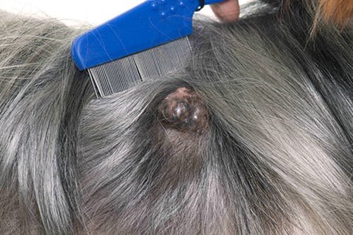
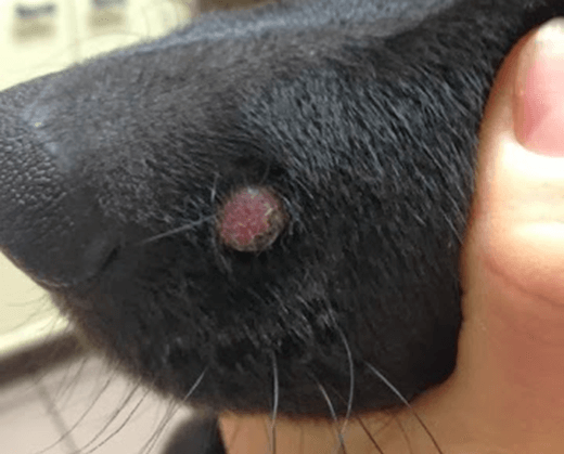
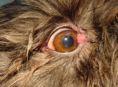
Histiocytoma in Dogs Pictures
Reference List
- Histiocytoma. https://www.epainassist.com/bones/what-is-fibrous-histiocytoma
- Radiation therapy. http://www.breastcancer.org/treatment/radiation/how_works
- Chemotherapy https://www.cancer.org/treatment/treatments-and-side-effects/treatment-types/chemotherapy/how-is-chemotherapy-used-to-treat-cancer.html
- Surgical excision. http://www.healthline.com/health/surgical-excision#purpose2
Similar Posts:
- Hemangiopericytoma
- Dermatofibroma
- Leiomyosarcoma – Survival Rate, Symptoms, Prognosis, Treatment
- Sebaceous Adenoma – Causes, Symptoms, Treatment, Pictures
- Chondrosarcoma – Symptoms, Prognosis, Treatment, Survival Rate
- Rhabdoid Tumor
- Tonsil Cancer – Pictures, Symptoms, Survival Rate, Staging, Prognosis


Leave a Reply