What is Gum Cancer?
Gum cancer is the malignancy of the soft tissue around the teeth, including the bones of the jaw underlying the teeth. Gum cancer is a type of oral cancer that affects predominantly males. It is also often called gingival cancer.
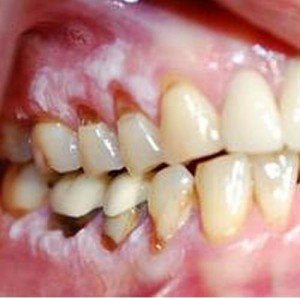
Gum Cancer showing Affectation of the Teeth
Gingival cancer can occur as a primary malignant tumor or as a metastatic site of other head and neck cancers. Gum cancer occurs in the form of squamous cell carcinoma, which means that the lining of the gums and mouth are affected.
Gum cancer is also highly metastatic because of being lymphoproliferative. This means that gum cancer cells are able to spread to other parts of the body through the lymphatic system. In fact, the most common site of metastasis is in the lymph nodes especially in the neck. Despite this, the progression of the disease is slow and early stages may be asymptomatic.
Signs & Symptoms of Gum Cancer
Signs and symptoms of gum cancer include those that affect the gums, teeth and oral cavity. These include:
- Pain in the gums
- Gingivitis or inflammation of the gums
- Tumor growth in the gums, (grows bigger overtime)
- Gum sores that do not heal
- Gum ulcerations
- Gum bleeding
- Discolorations in the gums (usually dark, reddish or white patches)
- Abnormal gum texture
- Loose teeth
Gum cancer can affect the teeth and may eventually cause complete absence of teeth. The roots of teeth can be affected, leading to dislocation, despite absence of cavities or damage. The earliest sign of gum cancer is gum bleeding. Often, people disregard this symptom thinking that it is just caused by trauma from brushing. However, gum bleeding in gingival cancer may occur, despite using a soft-bristled toothbrush. Smoking also masks discoloration of the gums caused by cancer since tar contained in cigarettes also causes gum discoloration.
Causes & Risk Factors of Gum Cancer
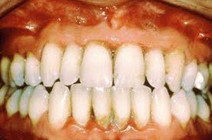
Periodontal Gum Disease as a Leading Cause of Gum Cancer
Causes and risk factors for gum cancer are similar to the general predisposing factors of oral cancer. These include:
Alcohol Intake
Presence of irritating chemicals in alcohol leads to changes in the cells of the gums. Alcohol produces drying effects on the gums, which leads to cellular dehydration. Chronic injury to the gums by chronic alcoholism produces abnormal cellular proliferation in an attempt to repair the injury, leading to tumor growth.
Smoking and Tobacco Use
Cigarette smoke and tobacco contains several carcinogenic chemicals that may lead to gum cancer. Smoking is attributed to most of the cases of oral cancer.
Betel Nut chewing
Certain populations of the world, such as Asian, have a traditional culture of chewing betel nut for dental purposes. However, dentists discourage the use of betel nut because it may lead to staining of the teeth, cavities, and even gingival and oral cancers.
Infection with Human Papilloma Virus (HPV)
Human Papilloma Virus may also spread to the mouth through unsafe sex practices. HPV produces cell changes in the gums and may lead to gingival cancer, aside from causing cervical and vulvar cancer.
Chronic inflammation and irritation
Chronic gingivitis and presence of periodontal abscess may also lead to gum cancer. Ill-fitting dentures, dental appliances and improper use of a toothbrush may also irritate the gums and cause cellular changes on the lining.
Male gender
The male population is at more risk for gum cancer because of lifestyle factors such as smoking and alcoholic beverage drinking, which are more commonplace and frequent among males.
Diagnosis
The diagnosis of gum cancer involves the following:
Medical History
A thorough medical history is done to assess previous diseases that may cause gum cancer. Risk factors are also assessed by determining the social and lifestyle history of the patient. Dietary and dental history is also assessed.
Physical examination
Physical examination of the gums is usually done by oral specialists or dentists. Examination includes thorough assessment of the gums and teeth including the whole oral cavity to determine any spread or underlying causes.
Biopsy
Physicians usually collect tissue samples from gingival ulcers and tumors and subject it to histologic examinations. Biopsy is the only test that determines malignancy in the gums.
X-ray
Dental X-ray and X-ray of the skull may also be performed to determine presence of tumors in the gums and other parts of the head.
Magnetic Resonance Imaging
Physicians may order further imaging tests such as MRI to have a more definite assessment of tumors and their location.
Treatment of Gum cancer
Treatment of gum cancer includes surgery, chemotherapy, radiation and other supportive managements.
Surgery
Surgery involves the removal of the gum tumor and is the first line of treatment for malignant tumors. Maxillectomy may be employed when the tumor has spread to the maxilla and palate. Radical neck dissection is also done when lymph node spread has occurred.
Chemotherapy
Chemotherapy is usually employed after surgery to kill cancer cells that have spread to other areas of the body. Combination drugs or single medication may be used. The administration of chemotherapy may potentially lead to bone marrow suppression because of destruction of normal cells, aside from the cancerous cells. Side-effects include anorexia, temporary hair loss, nausea, vomiting, and weight loss. Pre-chemotherapy drugs are also given, such as anti-emetics, to reduce side-effects of chemotherapy.
Radiation
Radiation therapy is also instituted as adjunct therapy to surgery to stop the spread and growth of malignant cells. Radiation is a localized management where radiation is directed on the tumor site.
Targeted Therapy
The use of cetuximab (Erbitux) targets cancerous cells specifically, which has fewer side-effects than chemotherapy. This drug targets the growth factor receptors, which prevents further tumor growth.
Rehabilitation Therapy
Patients with gum cancer should be assisted in returning to usual activities specifically, those involving eating and speaking. Patients should be taught to consume small frequent meals, consisting of a soft diet, to avoid undue tension in the teeth and gums. Speech therapy may also be employed to help the patient speak normally after surgery.
Reconstructive surgery may also be recommended to address disfiguring affects of gum and oral surgeries. Dental prosthesis is also available to restore the normal appearance of the gums, teeth, and mouth.
Prognosis and Survival Rate
Gum cancer has a good prognosis when detected at an early stage. Just like with any forms of cancer, distant metastasis provides the most unfavorable picture for patients. However, gum cancer has slow progression, which means that further spread of malignant cells is effectively prevented when treatments are instituted. The most common sites for metastases are the other parts of the oral cavity, lymph nodes, pharynx, larynx and thyroid gland. The mean 5 year survival rate of gum cancer is up to 70 to 80%, which is high when compared to other types of cancers.
Complications of Gum Cancer
Gum cancer also leads to complications resulting from affectation of other parts of the oral cavity and throat. Complications include:
- Dry mouth
- Poor salivation
- Impaired sensory function of the tongue, leading to difficulty in tasting foods
- Oral infections, such as lichen planus and candidiasis, due to a depressed immune system
- Difficulty forming speech because the gums also play a role in phonation
Gum cancers should be detected early in order for early management to be instituted. However, prevention is still the best key in managing gum cancers, which involves avoidance of the modifiable risk factors.
Gum Cancer Pictures
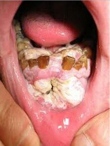
Advanced Gum Cancer Affecting the Rest of the Oral Cavity
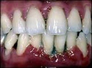
Localized Gum Cancer
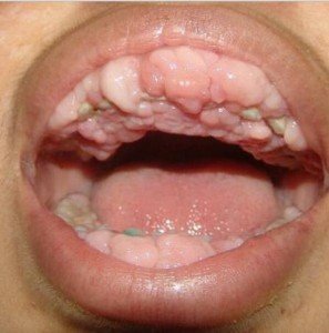
Gum Cancer Showing Gingival Hyperplasia or Inflammation
Staging
Gum cancer staging is based on oral cancer staging. Staging also involves either the extent of spread or the TNM staging system.
Oral cancer staging according to spread:
Stage I
Stage I gum cancer involves the growth of tumor, which is less than 2 cm in size. This stage does not involve lymph node spread yet.
Stage II
The tumor on the gum is more than 2 cm, but less than 4 cm in size. Still, there is no lymph node spread yet.
Stage III
Stage III gum cancer involves two developments. Either the tumor is more than 4 cm in size with no lymph node affectation or the tumor is less than 4 cm in size, but there is already spread to one lymph node. Tumors that have spread on to the lymph node are less than 3 cm in size.
Stage IV
The last stage of gum cancer involves spread of the tumor to the oral cavity and other adjacent tissues. There is also lymph node affectation of more than one lymph node, which may vary in size. There is also distant metastasis to other parts of the body.
TNM staging is also another way to characterize the stage of oral cancer. T stands for tumor; N stands for lymph nodes and M for distant metastasis.
Tumor
- TX- This stage describes that the primary tumor cannot be assessed yet
- T0- There is no indication of primary tumor
- Tis- Stands for carcinoma in situ
- T1- Less than 2cm diameter of tumor
- T2- Tumor is 2cm to 4cm in size
- T3- Tumor size is more than 4cm with spread to adjacent superficial tissues such as the tongue, sinus, etc.
- T4- Tumor spreads to the deep tissues of other organs in the oral cavity or other adjacent structures.
Lymph nodes
- NX- This stage describes that the regional lymph nodes cannot be assessed
- N0- No metastasis to lymph nodes
- N1- With metastasis to only one lymph node with 3cm or less in size
- N2- Metastasis to one or more lymph nodes not more than 6cm in size
- N3 Metastasis to lymph nodes with more than 6cm in size
Metastasis
- MX- Distant metastasis cannot be assessed yet
- M0- There is no distant metastasis
- M1- With distant metastasis
Proofreaded and updated by Alison on 22/10/2012
Similar Posts:
- Lip Cancer
- Tongue Cancer Pictures, Symptoms, Treatment, Prognosis, Survival Rate
- Vaginal Cancer – Symptoms, Signs, Pictures, Treatment, Causes
- Jaw Cancer
- Tonsil Cancer – Pictures, Symptoms, Survival Rate, Staging, Prognosis
- Duodenal Cancer – Symptoms, Prognosis, Survival Rate and Treatment
- Medullary Thyroid Cancer – Symptoms, Treatment, Prognosis



[…] Gum Cancer – Pictures, Symptoms, Signs, Treatment, Complications […]