A woman has its crown and glory. Some of them are proud of their hair, lips, eyes, nose, mouth, teeth, legs, hips, butt, body curves, skin complexion and most of their breast. The latter would be the sensitive one. The breast is consisting of fats, muscles and connective tissues. It is where new born latch on for breast milk. But what if the mother’s breast is affected by a disease condition? Can they still let their babies latch on for milk? What is this condition affecting it? Is there any treatment? Can they survive? Is it possible the baby can acquired this disease? What is the treatment for this condition? Where can we find support for the treatment? Is it fatal? What are the tests needed? Can the mother have higher survival rate? These are questions that frequently ask. We will answer it with the kind of disease we are going to tackle. This disease is Invasive Ductal Carcinoma (IDC).
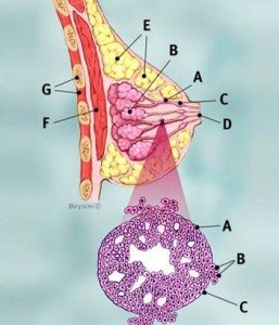
Invasive Ductal Carcinoma is the most common type of breast cancer. It is also known as Infiltrating Ductal Carcinoma. This carcinoma has damaged the wall of the milk ducts and started to conquer the breast tissues. It begins in the milk ducts that carry the milk-producing lobules in the nipples and goes deep down in the breast tissues. 80% of breast cancers are caused by this kind of carcinoma.

Each year about 180,000 women in the United States are affected and diagnosed with this type of cancer. Nonetheless, this affects women at early age but it more affected older women age 55 above. In rare cases it also affects men.
Invasive Ductal Carcinoma Signs and Symptoms
At first thought, women may not able to detect right away any changes in their breasts. However, when they look and feel for any mass or lumps in their breast which is not usual, they should take action immediately for it. They must do Breast Self-Examination (BES) to obtain this signs and symptoms. Look and feel for this:
- A mass or lumps in the breast
- Pain in the breast
- Skin dimpling around areola
- Having nipple discharges
- Pain in the nipple area
- Breast swelling
- Scaliness and redness in the nipple and skin in the breast
Tests and Procedures to Diagnose IDC
It’s hard to diagnose this condition alone. It requires a combination of procedures, examinations and specific testing.
- Breast mammography- this is an X-ray test to view the breast. The result of this shows finger-like projections and this projections shows invasion of the cancer cells.
- Ultrasound- also views additional tissues in the breast that affected by the cancer cells.
- Breast Physical Examination- note for any swelling, redness, scaliness or any unusual changes in the breast.
- Biopsy- this test will require some abnormal tissues to be taken out and do pathological test. There are two kinds of this biopsy:
- Core Need Biopsy- a large needle is inserted to the breast to extract samples from the breast and leaves a small scar in the breast. It has two types:
- Excisional biopsy- removes the entire suspiciously abnormal mass or lumps in the breast.
- Incisional biopsy- only small abnormal tissues is removed from the breast.
- Fine needle aspiration- a hollow and small needle is inserted in the breast and extract abnormal tissues. This procedure doesn’t leave a scar in the breast.
- Magnetic Resonance Imaging of the breast- it uses magnetic and radio waves thru a computer screen to view tissue of the breast. A dye is usually inserted to clearly view the abnormalities in the breast.
Invasive Ductal Carcinoma Cell Staging
Cancer has stages 1 to 4. In obtaining the right stages of the cancer cells, there are several factors to consider. These are:
- If the cancer cells has spread deep down the tissues
- If the cancer cells invades any parts of the body and adjacent lymph nodes
- The size and shape of the carcinoma
The grading of IDC:
- Grade 1- well differentiated or low grade
- Grade 2- moderately differentiated or intermediate stage
- Grade 3- poorly differentiated or high grade
Invasive Ductal Carcinoma Treatment
Listed below are the treatment modalities for invasive ductal carcinoma:
- Systemic Chemotherapy– generally used for malignant carcinoma.
Prognosis and Survival Rate
The prognosis depends on each patient’s factors like tumor size, histological considerations and biological factors. However, tumor that has bigger sizes are most likely with higher mortality rate. Tumor size that is 5 cm has 50-60% survival rate. With tumor sizes less than 1 cm has 90-98% plus 20 years survival rate.
- Mastectomy– removal of affected breast where the malignant carcinoma has invaded the breast tissues.
- Supportive care– after the success of the surgery, care is needed when providing wound dressing, changing of surgical patch and maintaining integrity of the skin.
- Continuous education– to be fully aware when the carcinoma regresses and the medication regimen already available.
Similar Posts:
- DCIS Breast Cancer
- Triple Negative Breast Cancer
- Intraductal Papilloma
- HER2 Positive Breast Cancer
- Urothelial Carcinoma
- Acinic Cell Carcinoma
- Top 40 Cancer Fighting Foods


Leave a Reply