What is mast cell tumor?
Mast cell neoplasms are a heterogeneous group of disorders that includes manifestations on the skin, like urticaria pigmentosa and systemic involvement, which affects all organ systems. It is a tumor of the immune system (also see other immune system tumors: chronic lymphocytic leukemia; Mycosis fungoides) This type of tumor is not well understood, because of the various functions of mast cells [1].
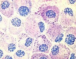
Mast cells
Mast cells are a part of immunological system. They are round cells with one nucleus. These cells are located in all tissue of human body in various quantities. They develop in bone marrow and spleen. Increased numbers of these cells are found in places where they can respond to antigens- skin, intestines, conjunctiva and airways. Mast cells are also found in heart tissue and various brain tissues like meninges and choroid plexus. These cells monitor blood for inflammation, therefore they are often found close to blood vessels, nerves and lymph. Mast cells produce biologically active compounds that can fight infection and also cause adverse effects.
Mast cells produce mediators in 3 different forms:
Preformed granules
These mediators that are released after mast cell activation
- Histamine
- Neutral proteases
- Heparin proteoglycans
Lipid mediators
- Prostaglandine D3
- Platelet activating factor

Cytokines and chemokines
- TNF-α
- Interleukin 4
All these compounds take a part in the immunological response against bacteria, viruses and parasites. But mast cells can also cause pathological immune response- mast cell activation disorders that include:
- Allergy
- Anaphylaxis
- Autoimmune diseases like rheumatoid arthritis
- Associated with antiphospholipid-antibody syndrome.
- Mast cell activation syndrome[2]
Causes
The causes of mast cell tumor are not known, but there are some factors that can trigger the disease, like use of NSAIDs, exposure to extreme temperatures or physical activity. Immune system is very complicated and the mechanisms are still being investigated [3].
Clinical manifestations of mast cell tumor
Indolent systemic mastocytosis
Clinically ISM manifests as:
- Hemodynamic instability
- Cutaneous mast cell disease
- Stomach and small intestine ulcers
- Malabsorbtions
- Infiltration of mast cells in intestines
- Infiltration in bone marrow

- Skeletal disease due to mast cell activity on bone surface
- Hepatosplenomegaly
- Lymphadenopathy (also see lip cancer)
Patients with ISM can live a normally long life, if the manifestations are appropriately managed[4].
Mastocytosis with associated clonal non-mast-cell lineage disease
Patients with this manifestation have abnormal amounts of mast cells in many organs and bone marrow abnormality, like myelodysplastic syndome or myeloproliferative disorder. The prognosis depends on the associated hematological disease.
Aggressive systemic mastocytosis
ASM is a rapidly progressing disease that starts out in the bone marrow and subsequently affects digestive tract, liver, spleen and lymph nodes.
Mast cell leukemia
MCL is very aggressive disease. It is characterized by increased amount of mast cells in bone marrow and blood. Survival rate is less than one year.
Mast cell sarcoma
MCS is very rare and aggressive neoplasm that usually presents as a solid mass [4,5].
Symptoms
The symptoms of mast cell tumor are hard to define and they depend on the organs involved in the process. Many patients experience unspecific systemic symptoms:
- Weakness
- Fatigue
- Sweating in the night
- Weight loss
- Depression
- Cognitive function decline
Some patients have described more specific symptoms:
- Attacks of flushing, sense of heat, heart palpitations and discomfort in chest, shortness of breath. These attacks sometimes are accompanied with nausea, vomiting, diarrhea, headache and feeling of light headedness.
- Syncope after stress, heat exposure, physical activities
- Flushing followed by decrease in blood pressure [4,5]
Cutaneous
Mast cell tumor can manifest in 4 different types of lesions:
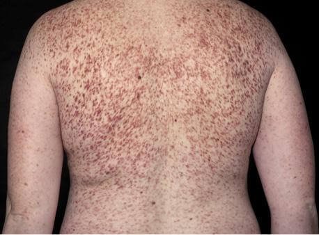
- Urticaria pigmentosa
- Red-brown macules, papules, plaques
- Randomly distributed
- Redness and edema can be present around lesions
- Blisters can form
- Telangiectasia macularis eruptive parstans
- Diffuse cuntaneous mastocytosis
- Rare, presents befor 3 years of age
- Involves al skin

- Skin has a yellow-brown color
- Dermatographism can be present
- Bleeding blisters are often present
- Mastocytoma
- Present at birth or appear within 3 months after birth
- Usually on extremities (also see: Dermatofibroma)
- Usually no systemic involvement[4,5]
Hematological
In systemic mast cell tumor usually bone marrow is involved. In this case symptoms are:
- Anemia
- Thrombocytopenia
- Leucopenia
- Leukocytosis
- Diffuse fibrosis of bone marrow and hypocellularity
Gastrointestinal
- Abdominal pain
- Diarrhea
- Nausea
- Vomiting
- Peptic ulcer
- Malabsorbtion
- Liver dysfunction, hepatomegaly and portal hypertension
- Splenomegaly
Other symptoms
- Lymphadenopathy-enlarged, painful lymphnodes (also see Acute lymphoblastic leukemia)
- Diffuse osteopenia with lesions in the bones causing pain and fractures [4,5]
Diagnosis
If mast cell tumor is suspected based on the clinical symptoms the following examinations should be done:
- Skin examination- gross appearance and microscopic evaluation of biopsy material
- Bone marrow biopsy
- Microscopic examination
- Immunohystochemistry
- Flow cytometry
- Blood analysis to measure serum tryptase levels ( ≥20 ng/ml)
- Bone scan
- Endoscopy and computed tomography of gastrointestinal tract[4]
Mast cell tumor is confirmed, when diagnostic criteria are met. Diagnostic criteria for cutaneous mastocytosis are: visible typical skin lesions of mast cell tumor and histological confirmation in skin biopsy. Systemic mast cell tumor is diagnosed based on major and minor criteria. Major criteria are:
- Dense mast cell infiltrates in bone marrow or other organ biopsy
- Infiltrates are confirmed using immunohistochemistry.
Minor criteria:
- Atypical morphology of more than 25% of mast cells- spindle shaped, immature or atypical
- Detection of KIT mutation in bone marrow, blood or other organs
- Mast cells that co-express CD117 with CD2
- Serum tryptase levels increased
The diagnosis can be made, if one major and on minor criteria are present or if 3 minor criteria are present [6].
Treatment
Treatment for mast cell tumor is usually symptomatic, but it does not alter the course of disease. One of the most important measures is to avoid triggering factors:
- Extreme temperatures
- Physical exertion
- Ethanol ingestion
- Use of NSAIDs
- Opiate analgesics
- Physical trauma
Medications
- Epinephrine- treatment of choice for hypotension
- Antihistamine prophylaxis to avoid episodes of hypotension, decrease symptoms of skin irritation and itchiness.
- H2 histamines- ranitidine, and proton pump inhibitors like omeprazole for treatment of gastritis and peptic ulcer
- Cromoglycolate- reduction of itchiness and formation of urtricaria
- Diphosphonates to treat osteopenia
- Topical corticosteroids to improve skin symptoms
- Systemic corticosteroids- decrease malabsorbtion and ascitis[7]
- Aspirin for episodes of syncope and flushing, but it can also worsen symptoms therefore aspirin should be used with caution[3, 4]
Other treatments
Radiation therapy can be used to limit localized mast cell tumor. Chemotherapy is used for mast cell tumors associated with myeloproliferative disease.
Splenectomy is used for patients with aggressive tumor, and it can limit cytopenia. Splenectomy can increase survival rate [4].
References
- What is mast cell tumor: http://link.springer.com/article/10.1007%2Fs12016-015-8487-6
- Mast cells: https://www.ncbi.nlm.nih.gov/pmc/articles/PMC3431027/
- Patient information: http://my.clevelandclinic.org/health/diseases_conditions/hic_Mastocytosis
- Symptoms and presentation: https://www.ncbi.nlm.nih.gov/pmc/articles/PMC1473141/
- Mast cell tumor symptoms, presentation, treatment http://www.nhs.uk/Conditions/Mastocytosis/Pages/Introduction.aspx#symptoms
- Diagnostic criteria: https://www.ncbi.nlm.nih.gov/pmc/articles/PMC1473141/table/t3/
- Cortisone effects: http://ebm.sagepub.com/content/80/4/677.short
Similar Posts:
- Granular Cell Tumor
- Squamous Cell Carcinoma in Situ – Pictures, Treatment, Symptoms
- Langerhans Cell Histiocytosis
- Chronic Myelogenous Leukemia
- Rhabdoid Tumor
- Chronic Lymphocytic Leukemia
- Childhood Leukemia


Leave a Reply