Rosettes are round groupings of cells that are found in tumors. There are different types of rosettes and it is vital to be familiar with each type because they assist in the diagnosis of difficult tumors [1, 2].
Rosettes
The round grouping of cells found in neurologic tumors are called rosettes because of their arrangements. These cells surround a central region that is similar to a spoke-wheel. They are named for their flower-like architecture that also similar to the rose windows installed in gothic cathedrals. The center of a rosette may be an empty-appearing lumen or a cytoplasmic process-filled space.
The cytoplasm of the cells in a rosette are wedge shaped and the nuclei of these cells are positioned peripherally. There are 4 main kinds of a rosette and each one is associated with a specific type of tumor.
The 4 types are Homer Wright rosette, Flexner-Wintersteiner rosette, True Ependymal rosette and Perivascular Pseudorosette. The mechanism that lead to the formation of these rosettes are not fully understood but their presence are associated with neuronal differentiation. It is important to note that these primary rosettes may only suggest a diagnosis and should be done in accordance with other tests in giving the definitive diagnosis [1, 2].
Homer Wright Rosette
This type of rosette is named after James Homer Wright. He is the first director of the Massachusetts General Hospital Pathology Laboratory and is also the developer of the Wright stain. He discovered these ball-like structures while he is studying adrenal and sympathetic nervous system malignancies that was later known as medulloblastomas. These rosettes enclose a meshwork of fibers that do not stain like the ones that are associated with neuroglia [1, 2]. Figure 1 shows an example of a Homer Wright Rosette.
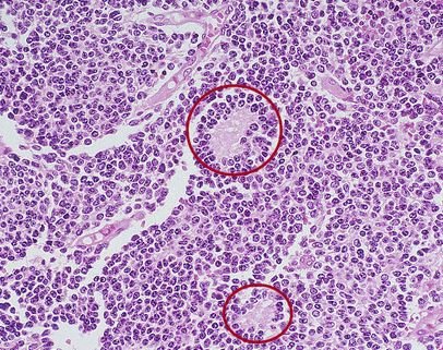
Figure 1- Homer Wright Rosette
The central area of a Homer Wright Rosette is made up of neuropil that contains primitive neuronal processes called neurites. It is hypothesized that the glycoproteins and glycolipids on the surface of the cells facilitate recognition of cell-to-cell and adhesion. The developing cells will then cluster together and their neurites will tangle. As these cells mature, their neurites stayed tangled in the center while their cell bodies are squeezed together in the peripheries thus forming a rosette [1, 2].
A Homer Wright rosette is commonly seen in the following neurologic tumors: medulloblastoma, olfactory neuroblastoma, primitive neruoectodermal tumors (PNET) and pineoblastoma. This kind of rosette can be seen in a posterior fossa tumor but this could be a pathognomonic sign of a medulloblastoma. The appearance of a Homer Wright Rosette may aid in establishing diagnosis but additional tests may be required for tumors that only has primitive-looking cells. These techniques include immunohistochemical analysis and special staining [1, 2, 3].
References
- Pathology Student. (2012, April 3). The four main types of rosettes in pathology. Retrieved from Pathology Student: http://www.pathologystudent.com/?p=5400
- Wippold, F. J., & Perry, A. (2006). Neuropathology for the Neuroradiologist: Rosettes and Pseudorosettes. American Journal of Neuroradiology, 488-492.
- Knipe, Henry, & Gerstenmaier, J. F. (2016). Homer Wright rosettes. Retrieved from Radiopaedia: http://radiopaedia.org/articles/homer-wright-rosettes
Similar Posts:
- Medulloblastoma
- Embryonal Rhabdomyosarcoma
- PNET Tumor
- Pineoblastoma
- Krukenberg Tumor
- Ganglioglioma
- Acute Myeloid Leukemia


Leave a Reply