Definition
Glomus tumor is a non-malignant vascular tumor that does not commonly occurs to a person and should not be confused with paraganglioma. The tumor arise from the glomus body which is a part of the skin’s dermis layer and is involved in the human body’s thermoregulation; as arterioles constricts with cold, glomus bodies expand in order to prevent heat loss.
The tumors are commonly noted in the foot, under the nail, or on the fingertip but can arise at other areas of the body. These growths are accountable for about less than 2% of all tumors in the soft tissue. The abnormal growths are tiny nodules of the distal extremities that are blue-red in color.
They commonly present in adults between 20-40 years of age. Most of the tumors are solitary and situated in cutaneous areas although other glomus tumors have extracutaneous involvement.
Glomus tumors are classified into three and these include:
- Solitary glomus tumors – A single tumor that exist in a specific area.
- Multiple glomus tumors – A number of tumors in specific areas.
- Congenital glomus tumors – Tumors that had been present since birth.
Symptoms of Glomus Tumor
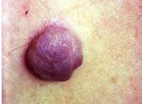
The common symptom of glomus tumor is the appearance of a reddish blue nodule that is about 1-2cm in diameter which is tender to touch. The small lesions may also become extremely painful that is described as bursting or burning.
The pain usually occurs due to a sudden change in temperature such as when the lesion is set in cold water. These benign growths may have a whitish appearance or a bluish discoloration. An elevation of the nail bed could also occur.
Although most cases of glomus tumors are benign, there are also malignant tumors which are mostly fatal. They are subdivided into three based on histologic appearance:
- Infiltrative glomus tumors (LIGT) – Contains the usual histologic appearance of the tumor
- Glomangiosarcomas arising in a benign glomus (GABG) – A cancerous tumor that arises and merges with the usual glomus tumor
- Glomangiosarcomas arising de novo (GADN) – It is the type of tumor that can hardly be recognized and must be differentiated from other kinds of round cell sarcomas
Causes
The exact reason why glomus tumors occur is not known. These tumors arise from neuromyoarterial plexus which is the modified smooth muscle cells of the glomus body. Glomus tumors are preferably thought of as hamartomas than true tumors.
A hamartoma is a non-cancerous tumorlike malformation composed of abnormal tissues and cells combined together as seen in certain parts of the body where growth occurs. The excruciating pain caused by the glomus tumors is also not fully understood although the pain neurotransmitter substance P in the nerve fibers had been spotted in the tumor.
Diagnosis
In imaging tests, glomus tumors appear as osteolytic lesions that are well restricted on particular areas. A sclerotic border is also present as a result of the slowly enlarging mass.
Diagnostic tests may involve:
Computed Tomography scan/CT scan
It is a special X-ray tests that produces cross-sectional pictures of the body. A non-specific subungual mass could be shown by a CT scan.
Conventional Radiography
It uses an imaging plate to create certain images. A bordered osseous thinning or erosion of the adjacent cortical bone is showed in the test.
Magnetic resonance imaging/MRI
The test can provide images of the body’s organs and structures. This is useful in detecting lesions in the soft tissues.
Ultrasound
Presence of subungal hypoechoic lesion with pain felt on the affected site. Hypervascularity is noted upon Doppler ultrasound.
A correct diagnosis is very essential for the treatment. Many of these lesions are most likely misdiagnosed as venous malformations or hemangiomas which make it even more difficult to come up with a precise assessment of the tumor.
Treatment
Treatment options for glomus tumor include:
Surgery – It involves the excision of tumors and this is the most preferred and recommended procedure to ease pain and lessen the chance of recurrence. The nail bed must be repaired after the subungual lesions have been removed.
Hypertonic saline
It is a saline solution that is injected to the patient.
Laser therapy
It is a therapy treatment that uses a strong beam of light for the tumors.
Sclerotherapy
It is a type of procedure that uses a certain medicine that is injected into the vessels.
Pictures
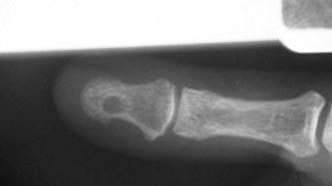
Picture 2 – X ray of Glomus Tumor
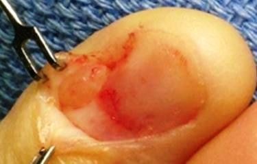
Image 3 – Surgical resection of glomus tumor
References:
- Glomus Tumor at http://www.bonetumor.org/tumors-vascular-tissue/glomus-tumor
- Glomus tumor Differential diagnosis, Treatment and prognosis, Radiographic features, Pathology at http://radiopaedia.org/articles/glomangioma
- http://www.dermnetnz.org/vascular/glomus.html
- Carlson ML, Sweeney AD, Wanna GB, Netterville JL, Haynes DS (2015 Jan). Natural history of glomus jugulare: a review of 16 tumors managed with primary observation. Otolaryngol Head Neck Surg. 152 (1):98-105.
- Jacob JT, Pollock BE, Carlson ML, Driscoll CL, Link MJ (2015 Jun). Stereotactic radiosurgery in the management of vestibular schwannoma and glomus jugulare: indications, techniques, and results. Otolaryngol Clin North Am. 48 (3):515-26.
- Forbes JA, Brock AA, Ghiassi M, Thompson RC, Haynes DS, Tsai BS (2012 Aug). Jugulotympanic paragangliomas: 75 years of evolution in understanding. Neurosurg Focus. 33(2):E13.
Similar Posts:
- Granular Cell Tumor
- Brenner Tumor
- Nerve Sheath Tumor
- Angiolipoma
- Solitary Fibrous Tumor
- Inoperable Brain Tumor
- Pilomatricoma – Treatment, Pictures, Symptoms, Causes, Prognosis


Leave a Reply