What is epithelioid sarcoma?
Epithelioid sarcoma (ES) has been a rarely developed malignancy affects soft tissues present in the upper limb. In vast description of ES attaining as a subcutaneous or dermal tissue mass in distal segments of the upper or lower limbs. This disease often affects young adults at their adolescent stage.
At the time of the first description about this type of sarcoma, medical science denotes this as “sarcoma aponeuroticum” due to its association with aponeuroses and adjacent structures. Clinician Enzinger first denoted this as “epithelioid sarcoma”.
Previously, this disease is often misdiagnosed due to epithelial and mesenchymal differentiation is not clearly detectable and at the initial stage the tumor progression, it may consider as chronic inflammatory processes, fibrohistiocytic tumor formation or necrotizing granulomas. The tumor progresses and extends all along with vascular channel lymph nodes and the lining of the ligaments, which leads to additional widespread inflammation and a primitive mass. (1,2)
Sign and symptoms
The appearance of tumor/ lump seems as tiny, solid nodules sited in the superficial or deep, soft tissues and can develop in single or cluster mode. Local multifocal concord is an atypical trait exhibit by this lump. This lump is often wrongly diagnosed as a cutaneous condition, corns or warts,, and a proper identification may be overdue with severe clinical presentation.
A maximum number of the tumor is not painful. The tumor can affect both superficial and deeper layers of the tissues.
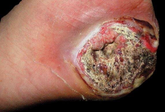
In case of superficially located tumor, it forms a solid lump that may present as single or multiple cluster with thick covered and demonstrated as a ” firm lump” or “woody hard knot”. This type of tumor is usually painless and slowly progressive. Deep sited Tumors are also formed nodules and often seem as skin elevation at the initial stage, but gradually becomes ulcerated after some weeks or months.
Frequently such lacerations are misdiagnosed as a “draining abscess”,” indurated ulcer”, or “infected wart” , which does heal with conventional therapy used to treat the condition. The shape of the tumors is encircled with having a range of varying size, which may lie in between 3 -6 cm in diameter.
According to the following findings from different case studies are considered as common symptoms in epithelioid sarcoma :
- Firm tiny mass
- May be solitary or cluster lump formation on the skin
- Distal part of the upper and lower limbs, such as fingers, toes, hands, feet and forearm are common affected sites
- The mass is hard and firm to the touch (location – dermis/deep soft tissue)
- Superficial mass usually turn to ulcerate
- Rarely, pain and swelling occurs
- If underlying nerve gets affected then regional weakness or numbness also occur.
- Lymph nodes, lung and scalp is often affected due to metastasis (1,2,3,5)
Diagnosis
Epithelioid sarcoma is diagnosed by following methods
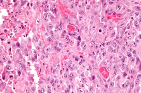
Biopsy
The small section of tissue is collected from the mass for biopsy test and often provide confirmatory result.
Affected lymph node biopsy
To this is often recommended to check the metastasis of the epithelioid sarcoma with affected lymph node involvement.
Staining of immuno-histochemical
This test helps to identify the involve gene and mutagenic changes which influence the epithelioid sarcoma.
MRI
Doctors usually order an MRI for detecting location or site of the mass formation and this is a primary test, which guide clinicians the requirement of performing the biopsy and other the pathologic analysis. After the surgical intervention, The MRI may also conduct for determining the possibility of the recurrence of the tumor. (3,5)

Epitheliod Sarcoma MRI Forearm
Treatment
Early and correct diagnosis is the important factor for treating this type of sarcoma. Before conducting the treatment plan, it is also crucial to know the extension of the tumor and the organs involved in the epithelioid sarcoma.
Usually surgical removal of the affected local lymph node or nodes is adapted by the clinician to restrict the metastasis. Often amputation of toe or finger is commonly required if the tumor is only extended in the toe or finger.
Limited treatment options are available for highly metastatic stage. Conservation of a functional limb exclusion of using the prosthesis. Sometimes the whole tumor cannot be removable due to its location, then may doctor prefer intra-lesional or marginal scraping.
Local and regional spreading of sarcoma can also treated by amputation. Amputation is mainly preferred for restricting the recurrence, though the chance is not limited to this process in every case. If the metastasis already spread to outlying site, then amputation cannot be beneficial to stop the recurrence.
Surgical therapy often combined with adjuvant therapy, including chemotherapy or radiotherapy, as the sarcoma is highly metastatic. Doxorubicin is the preferable choice as chemotherapeutic agent. (3,4,5)
Survival report and Prognosis
Research report revealed that the survival rate is 5 years maximum in 60% to 75% cases with epithelioid sarcoma. But this data is controversial, as this rate is improved in case of early, accurate detection with correct surgical and adjuvant therapy applied to younger aged people.
In more specific cases also denoted that females provide superior prognosis rate than the males. The negative prognosis of the treatment and survival rate linked with the size of the tumor.
It has been observed that more than 2 cm diameter of the tumor can cause inferior outcome of the treatment and survival rate become less, as this often affect the vascular tissue and wide metastasis can involve vital organs. Therefore, vascular invasion is a very important factor for better survival and positive prognosis. (4,5)
References
1. Joseph F. Sobanko, Lindsay Meijer, and Thomas P. Nigra, (2009); Epithelioid Sarcoma; J Clin Aesthet Dermatol. 2009 May; 2(5): 49–54; Retrieve from: http://www.ncbi.nlm.nih.gov/pmc/articles/PMC2924131/
2. Tadashi Hasegawa, Yoshihiro Matsuno, Tadakazu Shimoda, Toru Umeda, Ryohei Yokoyama and Setsuo Hirohashi, (2001); Proximal-Type Epithelioid Sarcoma: A Clinicopathologic Study of 20 Cases; Mod Pathol 2001;14(7):655–663; Retrieve from: http://www.nature.com/modpathol/journal/v14/n7/full/3880368a.html
3. Epithelioid Sarcoma; Retrieve from: http://www.bonetumor.org/tumors-fibrous-tissue/epithelioid-sarcoma
4. Muhammad Umar Jawad, Jason Extein, Elijah S. Min, and Sean P. Scully; Prognostic Factors for Survival in Patients with Epithelioid Sarcoma: 441 Cases from the SEER Database; Clin Orthop Relat Res. 2009 Nov; 467(11): 2939–2948; Retrieve from: http://www.ncbi.nlm.nih.gov/pmc/articles/PMC2758965/
5. Epithelioid Sarcoma; Retrieve from: http://healthh.com/epithelioid-sarcoma/
Similar Posts:
- Clear Cell Sarcoma
- Ewing’s Sarcoma – Prognosis, Survival Rate, Symptoms, Treatment
- Malignant Mesothelioma
- Angiosarcoma – Prognosis, Symptoms, Treatment, Pictures, Causes
- Leiomyosarcoma
- Embryonal Rhabdomyosarcoma
- Leiomyosarcoma – Survival Rate, Symptoms, Prognosis, Treatment


Leave a Reply