What is Angiolipoma?
Angiolipoma is a subcutaneous benign tumor that mainly consists of fatty tissue and blood vessels. It belongs to a group of benign tumors called lipomas. In some cases angiolipoma can be present in muscle tissue. This benign tumor can occur at any age or gender, but it is most commonly seen in young adults[1].
Causes
The exact cause of angiolipoma is not known. It has been observed, that in some cases this tumor seems to run in families[1].
Symptoms
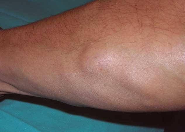
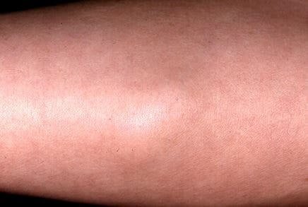
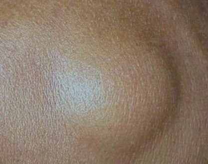
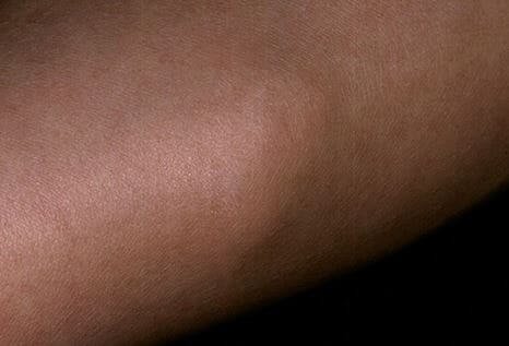
Angiolipoma can present as a single nodule in one or many locations. Usually 2-4 angiolipomas are present. Most typical location is on the forearm. It can also be located on the arm, chest, back or lower leg area. These tumors are rarely present in the head and neck area. Angiolipoma usually has well circumscribed borders. The nodule is often painful, but in some cases it can only present with no symptoms. Angiolipoma is rarely larger than 2cm in diameter.
In some cases angiolipoma can cause severe pain. If it is big in size, it can also damage nerves and blood vessels[2]. Also see glomus tumor.
Diagnosis
The diagnosis is usually made by obtaining thorough patient history, examining the lesion and performing tissue biopsy. Patients usually are 20 to 30 years old and complain about slow growing lesion, that causes pain. Sometimes the lesion is painful only when palpating, but in some cases it causes no pain. It is usually located on the forearm and not bigger than 2cm. There are no blood tests that can confirm the diagnosis. Usually biopsy is obtained to confirm diagnosis [1,6].
Histology
Angiolipoma is encapsulated and composed of mature fat cells and small blood vessels. The blood vessels can have patchy distribution, frequently they are accentuated in the subcapsular area. Most of them are capillaries with thick walls. One of the most important diagnostic features is the presence of fibrin thrombi. In older lesions fibrosis can be observed. Mast cells can be present.
In immunohistochemistry, the blood vessels cells (endothelium) are positive for CD34 and CD31 markers. Genetic testing is usually normal [2,3].

 Imaging studies
Imaging studies of subcutaneous lesions usually have no diagnostic significance, but in some cases magnetic resonance or ultrasound can be performed. In both of these investigations a well circumscribed lesion containing fat tissue and vascular component can be seen[2,3].
Types
Infiltrative angiolipoma
In some cases tumors containing fat tissue and blood vessels were found. These were previously considered to be infiltrative angiolipomas, but currently they are known as intramuscular hemangiomas. These lesions mostly consist of blood vessels but fat cells are also present. Infiltrative angiolipoma is usually found in muscle tissue. These lesions are encapsulated. In some cases it can cause neural deficits due to pressure on nerves or blood vessels[4].
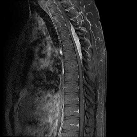
Spinal angiolipoma
Angiolipoma can also present as benign spinal tumor. Although it is not a spinal cancer, presence of angiolipoma can cause a variety of symptoms. This neoplasm is extremely rare, with only around 100 known cases. They are more commonly present in women between 50 to 60 years of age. Although spinal angiolipomas are slow growing lesions, they can cause severe symptoms due to spinal cord compression. Symptoms vary depending on the location of the tumor. Most commonly they are located in the thoracic spine. Symptoms include:
- Paresis under the level of lesion
- Pain in the back
- Sensory changes
- Hyper- reflexia in lower limbs[5]
![]()
Differential Diagnosis
- Kaposi sarcoma– malignant disease associated with HIV infection. Presents as patches, plaques or nodules that can be similar to angiolipoma. Histological investigation reveals cytologic atypia with red cell extravasion, which is not a typical finding in angiolipoma.
- Spindle cell hemangioendothelioma is a benign tumor composed of blood vessels. In comparison with angiolipoma, SCH contains more cavernous vessels and is poorly localized.
- Angioleiomyoma- angioleiomyoma sometimes has a fat component present therefore it should be differenciated from angiolipoma.
- Angiomyolipoma- angiomyolipoma is also a type of lipoma. Besides fat tissue and blood vessels it also has muscle tissue present. Immunohistochemistry reveals positive antibody- HMB-45, which is not present in angiolipoma[3].
Treatment
If angiolipoma presents with no symptoms, it can be left with no treatment. Most commonly it causes pain or discomfort and the method of choice is surgical excision. Usually the whole tumor is removed and it rarely reoccurs. If some parts of tumor are not removed, angiolipoma is likely to recur. This tumor is not likely to become malignant (also read: angiosarcoma).[6]
If you found this article helpful, share it on social media. For your personal experience, use the comments box below.
Pictures
References
- General information: http://www.dovemed.com/diseases-conditions/angiolipoma/
- Presentation: http://www.pathologyoutlines.com/topic/softtissueadiposeangiolipoma.html
- Histology and differential diagnosis: http://surgpathcriteria.stanford.edu/softfat/lipoma/angiolipoma.html
- Infiltrative angiolipoma: http://www.ijss-sn.com/uploads/2/0/1/5/20153321/ijss_dec_cr04_20150102_v2.pdf
- Spinal angiolipoma: https://www.ncbi.nlm.nih.gov/pmc/articles/PMC2565558/
- Treatment: http://www.primehealthchannel.com/angiolipoma-definition-causes-symptoms-and-treatment.html
Similar Posts:
- Squamous Cell Carcinoma in Situ – Pictures, Treatment, Symptoms
- Granular Cell Tumor
- Keratoacanthoma – Pictures, Treatment, Symptoms, Causes
- Spinal Cancer – Symptoms, Prognosis, Survival Rate, Life Expectancy
- Langerhans Cell Histiocytosis
- Angiosarcoma – Prognosis, Symptoms, Treatment, Pictures, Causes
- Urethral Cancer


Leave a Reply